
Diagnostic imaging, often referred to as diag image, has dramatically transformed the landscape of healthcare. These non-invasive technologies allow medical professionals to peer inside the human body, offering a deeper understanding of a patient’s health status. By providing high-resolution images, diag imaging helps clinicians diagnose conditions, plan treatments, and monitor diseases with greater precision, all without the need for invasive procedures. Over the years, technologies such as X-rays, CT scans, MRI, ultrasound, and PET scans have become integral to modern medical practice.
What is Diagnostic Imaging?
At its core, diagnostic imaging is a group of technologies used to create detailed visual representations of the internal structures and functions of the human body. These images aid physicians in diagnosing medical conditions, tracking the progress of diseases, and guiding treatment plans. Diagnostic imaging covers a broad range of methods, each with its own strengths and applications. The common denominator, however, is the ability to view and assess the body’s internal workings without surgery or other invasive measures.
From the initial discovery of X-rays to today’s sophisticated modalities, diagnostic imaging has evolved to offer greater insight into various conditions, from broken bones to complex neurological disorders. This suite of techniques is now indispensable in the medical field, allowing for quicker, more accurate diagnoses and better outcomes for patients.
The Evolution of Diagnostic Imaging
The first major breakthrough in diagnostic imaging occurred in 1895 with the discovery of X-rays by Wilhelm Conrad Roentgen. Initially, X-rays were used primarily for identifying fractures and other issues related to bone structure. Over time, the technology improved, and with it, the types of conditions that could be diagnosed using X-rays expanded. In the early years, X-ray images were captured on photographic film, which was a tedious process involving physical development of the images.
The shift to digital imaging, which took place in the late 20th and early 21st centuries, marked another significant milestone in the evolution of diagnostic imaging. Digital systems allow for clearer, more detailed images, which can be manipulated, stored, and shared instantly. This shift also significantly improved the speed of diagnoses, as doctors no longer had to wait for films to be processed. As a result, digital imaging systems have become integral to modern hospitals and clinics, with over 85% of hospitals worldwide utilizing these technologies as part of their electronic health record (EHR) systems.
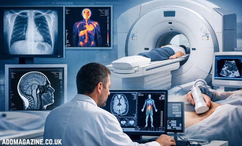
Key Modalities in Diagnostic Imaging
There are several types of diagnostic imaging technologies in use today, each serving distinct purposes based on the type of information required by healthcare professionals.
1. X-ray Imaging
X-ray imaging is perhaps the most well-known diagnostic tool. It uses ionizing radiation to capture images of dense structures within the body, such as bones. While its primary application is in the detection of fractures and bone-related issues, it is also useful in identifying conditions in other areas such as the lungs, chest, and abdominal region. For example, chest X-rays are commonly used to detect lung infections or conditions such as pneumonia or tuberculosis. X-ray machines are fast, affordable, and widely accessible, making them an essential tool in emergency rooms and clinics around the world.
2. Computed Tomography (CT) Scans
CT scans, or computed tomography scans, are more advanced than traditional X-rays. They use multiple X-ray images taken from different angles and combine them to produce cross-sectional images of the body. These images provide a detailed view of the body’s internal structures, including organs, tissues, and blood vessels. Unlike X-rays, which provide flat images, CT scans create three-dimensional representations, making them especially useful for diagnosing complex conditions.
CT scans are particularly valuable in the diagnosis of conditions such as:
- Cancer
- Heart disease
- Internal injuries or bleeding
- Neurological conditions such as stroke
The high-resolution images generated by CT scans allow healthcare providers to detect problems at earlier stages and monitor the progress of treatments over time.
3. Magnetic Resonance Imaging (MRI)
MRI uses magnetic fields and radio waves to create detailed images of the body’s internal structures. Unlike X-rays and CT scans, which rely on ionizing radiation, MRI is considered a safer alternative, as it does not involve radiation exposure. MRI is especially useful for imaging soft tissues, such as the brain, spinal cord, muscles, and organs. It has become one of the most important diagnostic tools for a wide range of medical conditions.
The demand for MRI has increased significantly in recent years. In fact, MRI usage has risen by around 50% globally over the last decade. This increase highlights the technology’s critical role in diagnosing neurological disorders like multiple sclerosis and brain tumors, as well as spinal conditions such as herniated discs, and musculoskeletal issues including ligament tears and joint injuries.
4. Ultrasound
Ultrasound imaging, also known as sonography, utilizes high-frequency sound waves to produce images of internal structures in real-time. This technology is particularly useful for monitoring pregnancies, diagnosing abdominal conditions, and evaluating organs like the liver, kidneys, and heart. Ultrasound is non-invasive, safe, and does not involve radiation, which makes it a preferred option for pregnant women and for imaging soft tissues. For those who require faster results or a more personalized diagnostic experience, booking a private ultrasound scan near me can offer immediate peace of mind without the long waiting lists often found in traditional healthcare settings.
Some common applications of ultrasound include:
- Monitoring fetal development during pregnancy
- Diagnosing gallstones or kidney stones
- Evaluating the heart’s function
- Assessing blood flow in veins and arteries
Ultrasound is also used in guiding certain medical procedures, such as biopsies or injections.
5. Positron Emission Tomography (PET) Scans
PET scans involve the injection of a small amount of radioactive material into the body, which accumulates in metabolically active tissues. The scanner then detects the radiation and produces detailed images of these areas. PET scans are primarily used to evaluate the metabolic activity of tissues, making them particularly useful in cancer detection and monitoring. And PET scans help to determine the size, shape, and location of tumors, as well as how the tumors are responding to treatments.
PET scans are also used in the diagnosis of neurological conditions such as Alzheimer’s disease, where they help detect changes in brain activity. These scans play an integral role in staging cancer, identifying metastasis (spread of cancer), and assessing the effectiveness of treatment regimens.
The Role of Artificial Intelligence in Diagnostic Imaging
Artificial intelligence (AI) is rapidly reshaping the field of diagnostic imaging. AI algorithms can now analyze medical images with remarkable speed and accuracy, assisting radiologists in detecting abnormalities, interpreting results, and reducing the likelihood of human error. AI has shown potential in enhancing the precision of diagnoses, from identifying subtle patterns that may be missed by the human eye to automating certain routine tasks like image categorization.
Some ways in which AI is revolutionizing diagnostic imaging include:
- Faster Image Interpretation: AI algorithms can quickly process and interpret medical images, which can help in urgent care settings where time is critical.
- Improved Detection Rates: AI tools are able to recognize patterns that might be too subtle for human radiologists, increasing the likelihood of early disease detection.
- Enhanced Workflow Efficiency: AI systems can automate parts of the diagnostic process, streamlining tasks and allowing healthcare providers to focus on patient care.
In the coming years, AI is expected to become more integrated into diagnostic imaging, improving workflows, reducing errors, and enabling more personalized care for patients.
The Future of Diagnostic Imaging
The future of diagnostic imaging looks promising, with ongoing advancements in technology, AI, and machine learning. The next frontier includes improving image resolution, reducing scan times, and making these technologies more widely accessible, especially in underserved or rural areas. The integration of real-time imaging data into healthcare systems, coupled with AI-driven insights, will continue to drive improvements in diagnosis, treatment planning, and patient care.
Furthermore, as personalized medicine continues to gain ground, diagnostic imaging will play a pivotal role in tailoring treatments to individual patients. With the ability to analyze genetic, molecular, and imaging data, clinicians will be able to make more informed decisions that are specific to each patient’s unique needs.
Conclusion
Diagnostic imaging is a cornerstone of modern medicine, providing clinicians with the tools they need to diagnose, treat, and monitor a wide range of medical conditions. From the early days of X-rays to the advanced imaging modalities used today, diag imaging has continually evolved, offering increasingly detailed and accurate representations of the human body. With the integration of AI and other emerging technologies, the field of diagnostic imaging is set to continue its transformative impact on healthcare, improving outcomes for patients worldwide.